Pregnancy is the carrying of one or more offspring, known as a fetus or embryo, in the womb of a woman. In a pregnancy, there can be multiple gestations, as in the case of twins or triplets. Childbirth usually occurs about 38 weeks after conception; in women who have a menstrual cycle length of four weeks, this is approximately 40 weeks from the last normal menstrual period (LNMP). The World Health Organization defines normal term for delivery as between 37 weeks and 42 weeks. Human pregnancy is the most studied of all mammalian pregnancies.
One scientific term for the state of pregnancy is gravidity (adjective "gravid"), latin for “heavy” and a pregnant female is sometimes referred to as a gravida.[1] Similarly, the term parity (abbreviated as “para”) is used for the number of previous successful live births. Medically, a woman who has never been pregnant is referred to as a “nulligravida”, a woman who is (or has been only) pregnant for the first time as a “primigravida”,[2] and a woman in subsequent pregnancies as a multigravida or “multiparous.”[1][3][4] Hence, during a second pregnancy a woman would be described as “gravida 2, para 1” and upon live delivery as “gravida 2, para 2.” An in-progress pregnancy, as well as abortions, miscarriages, or stillbirths account for parity values being less than the gravida number. In the case of twins, triplets etc., gravida number and parity value are increased by one only. Women who have never carried a pregnancy achieving more than 20 weeks of gestation age are referred to as “nulliparous.”[5]
The term embryo is used to describe the developing offspring during the first 8 weeks following conception, and the term fetus is used from about 2 months of development until birth.[6][7]
In many societies’ medical or legal definitions, human pregnancy is somewhat arbitrarily divided into three trimester periods, as a means to simplify reference to the different stages of prenatal development. The first trimester carries the highest risk of miscarriage (natural death of embryo or fetus). During the second trimester, the development of the fetus can be more easily monitored and diagnosed. The beginning of the third trimester often approximates the point of viability, or the ability of the fetus to survive, with or without medical help, outside of the uterus.[8]
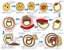
 The initial stages of human embryogenesis.Although pregnancy begins with implantation, the process leading to pregnancy occurs earlier as the result of the female gamete, or oocyte, merging with the male gamete, spermatozoon. In medicine this process is referred to as fertilization; in lay terms, it is more commonly known as “conception.” After the point of fertilization, the fused product of the female and male gamete is referred to as a zygote or fertilized egg. The fusion of male and female gametes usually occurs following the act of sexual intercourse, resulting in spontaneous pregnancy. However, the advent of artificial insemination and in vitro fertilisation have also made achieving pregnancy possible in cases where sexual intercourse does not result in fertilization (e.g., through choice or male/female infertility).
The process of fertilization occurs in several steps, and the interruption of any of them can lead to failure. Through fertilization, the egg is activated to begin its developmental program, and the haploid nuclei of the two gametes come together to form the genome of a new diploid organism [9]
At the beginning of the process, the sperm undergoes a series of changes. As freshly ejaculated sperm is unable or poorly able to fertilize,[10] it must undergo capacitation in the female's reproductive tract over several hours. This increases its motility and destabilizes its membrane, preparing it for the acrosome reaction, the enzymatic penetration of the egg's tough membrane, the zona pellucida, which surrounds the oocyte.
Legal regulations in different countries include gestation age beginning from 16 to 22 weeks (5 months) before birth.
There is a standard deviation of 8-9 days surrounding due dates calculated with even the most accurate methods. This means that fewer than 5 percent of births occur at exactly 40 weeks; 50 percent of births are within a week of this duration, and about 80 percent are within 2 weeks.[13] It is much more useful and accurate, therefore, to consider a range of due dates, rather than one specific day, with some online due date calculators providing this information. [2]
Pregnancy is considered "at term" when gestation attains 37 complete weeks but is less than 42 (between 259 and 294 days since LMP). Events before completion of 37 weeks (259 days) are considered preterm; from week 42 (294 days) events are considered postterm.[14] When a pregnancy exceeds 42 weeks (294 days), the risk of complications for both the woman and the fetus increases significantly.[12][15] Therefore, in an otherwise uncomplicated pregnancy, obstetricians usually prefer to induce labour at some stage between 41 and 42 weeks.[16][17]
Birth before 39 weeks, even if considered "at term", increases the risk of complications and premature death, from factors including under-developed lungs, infection due to under-developed immune system, problems feeding due to under-developed brain, and jaundice from under-developed liver. Some hospitals in the United States have noted a significant increase in neonatal intensive care unit patients when women schedule deliveries for convenience and are taking steps to reduce induction for non-medical reasons.[18] Complications from Caesarean section are more common than for live births.
Recent medical literature prefers the terminology preterm and postterm to premature and postmature. Preterm and postterm are unambiguously defined as above, whereas premature and postmature have historical meaning and relate more to the infant's size and state of development rather than to the stage of pregnancy.[19][20]
Accurate dating of pregnancy is important, because it is used in calculating the results of various prenatal tests, (for example, in the triple test). A decision may be made to induce labour if a fetus is perceived to be overdue. Furthermore, if LMP and ultrasound dating predict different respective due dates, with the latter being later, this might signify slowed fetal growth and therefore require closer review.
The age of fetal viability has been receding because of continued medical progress. Whereas it used to be 28 weeks, it has been brought back to as early as 23, or even 22 weeks in some countries.[citation needed]
A woman is considered to be in labour when she begins experiencing regular uterine contractions, accompanied by changes of her cervix — primarily effacement and dilation. While childbirth is widely experienced as painful, some women do report painless labours, while others find that concentrating on the birth helps to quicken labour and lessen the sensations. Most births are successful vaginal births, but sometimes complications arise and a woman may undergo a cesarean section.
During the time immediately after birth, both the mother and the baby are hormonally cued to bond, the mother through the release of oxytocin, a hormone also released during breastfeeding.
Most pregnant women experience a number of symptoms,[21] which can signify pregnancy. The symptoms can include nausea and vomiting, excessive tiredness and fatigue, cravings for certain foods that are not normally sought out, and frequent urination particularly during the night.
A number of early medical signs are associated with pregnancy.[22][23] These signs typically appear, if at all, within the first few weeks after conception. Although not all of these signs are universally present, nor are all of them diagnostic by themselves, taken together they make a presumptive diagnosis of pregnancy. These signs include the presence of human chorionic gonadotropin (hCG) in the blood and urine, missed menstrual period, implantation bleeding that occurs at implantation of the embryo in the uterus during the third or fourth week after last menstrual period, increased basal body temperature sustained for over 2 weeks after ovulation, Chadwick's sign (darkening of the cervix, vagina, and vulva), Goodell's sign (softening of the vaginal portion of the cervix), Hegar's sign (softening of the uterus isthmus), and pigmentation of linea alba – Linea nigra, (darkening of the skin in a midline of the abdomen, caused by hyperpigmentation resulting from hormonal changes, usually appearing around the middle of pregnancy).[22][23] Breast tenderness is common during the first trimester, and is more common in women who are pregnant at a young age.[24]
Pregnancy detection can be accomplished using one or more various pregnancy tests,[25] which detect hormones generated by the newly formed placenta. Clinical blood and urine tests can detect pregnancy 12 days after implantation.[26] Blood pregnancy tests are more accurate than urine tests.[27] Home pregnancy tests are urine tests, and normally cannot detect a pregnancy until at least 12 to 15 days after fertilization. A quantitative blood test can determine approximately the date the embryo was conceived.
In the post-implantation phase, the blastocyst secretes a hormone named human chorionic gonadotropin, which in turn stimulates the corpus luteum in the woman's ovary to continue producing progesterone. This acts to maintain the lining of the uterus so that the embryo will continue to be nourished. The glands in the lining of the uterus will swell in response to the blastocyst, and capillaries will be stimulated to grow in that region. This allows the blastocyst to receive vital nutrients from the woman.
Despite all the signs, some women may not realize they are pregnant until they are quite far along in their pregnancy. In some cases, a few women have not been aware of their pregnancy until they begin labour. This can be caused by many factors, including irregular periods (quite common in teenagers), certain medications (not related to conceiving children), and obese women who disregard their weight gain. Others may be in denial of their situation.
An early obstetric ultrasonography can determine the age of the pregnancy fairly accurately. In practice, doctors typically express the age of a pregnancy (i.e., an "age" for an embryo) in terms of "menstrual date" based on the first day of a woman's last menstrual period, as the woman reports it. Unless a woman's recent sexual activity has been limited, she has been charting her cycles, or the conception is the result of some types of fertility treatment (such as IUI or IVF), the exact date of fertilization is unknown. Without symptoms such as morning sickness, often the only visible sign of a pregnancy is an interruption of the woman's normal monthly menstruation cycle, (i.e., a "late period"). Hence, the "menstrual date" is simply a common educated estimate for the age of a fetus, which is an average of 2 weeks later than the first day of the woman's last menstrual period. The term "conception date" may sometimes be used when that date is more certain, though even medical professionals can be imprecise with their use of the two distinct terms. The due date can be calculated by using Naegele's rule. The expected date of delivery may also be calculated from sonogram measurement of the fetus. This method is slightly more accurate than methods based on LMP.[28] Additional obstetric diagnostic techniques can estimate the health and presence or absence of congenital diseases at an early stage.
Diagnostic criteria are: Women who have menstrual cycles and are sexually active, a period delayed by a few days or weeks is suggestive of pregnancy; elevated B-hcG to around 100,000 mIU/mL by 10 weeks of gestation
Morning sickness occurs in about seventy percent of all pregnant women, and typically improves after the first trimester.[31][dead link] Although described as "morning sickness", women can experience this nausea during afternoon, evening, and throughout the entire day.
Shortly after conception, the nipples and areolas begin to darken due to a temporary increase in hormones.[32] This process continues throughout the pregnancy.
The first 12 weeks of pregnancy are considered to make up the first trimester. The first two weeks from the first trimester are calculated as the first two weeks of pregnancy even though the pregnancy does not actually exist. These two weeks are the two weeks before conception and include the woman's last period.
The third week is the week in which fertilization occurs and the 4th week is the period when implantation takes place. In the 4th week, the fecundated egg reaches the uterus and burrows into its wall which provides it with the nutrients it needs. At this point, the zygote becomes a blastocyst and the placenta starts to form. Moreover, most of the pregnancy tests may detect a pregnancy beginning with this week.
The 5th week marks the start of the embryonic period. This is when the embryo's brain, spinal cord, heart and other organs begin to form.[33] At this point the embryo is made up of three layers, of which the top one (called the ectoderm) will give rise to the embryo's outermost layer of skin, central and peripheral nervous systems, eyes, inner ear, and many connective tissues.[33] The heart and the beginning of the circulatory system as well as the bones, muscles and kidneys are made up from the mesoderm (the middle layer). The inner layer of the embryo will serve as the starting point for the development of the lungs, intestine and bladder. This layer is referred to as the endoderm. An embryo at 5 weeks is normally between 1⁄16 and 1⁄8 inch (1.6 and 3.2 mm) in length.
In the 6th week, the embryo will be developing basic facial features and its arms and legs start to grow. At this point, the embryo is usually no longer than 1⁄6 to 1⁄4 inch (4.2 to 6.3 mm). In the following week, the brain, face and arms and legs quickly develop. In the 8th week, the embryo starts moving and in the next 3 weeks, the embryo's toes, neck and genitals develop as well. According to the American Pregnancy Association, by the end of the first trimester, the fetus will be about 3 inches (76 mm) long and will weigh approximately 1 ounce (28 g).[34]
The uterus, the muscular organ that holds the developing fetus, can expand up to 20 times its normal size during pregnancy.
Although the fetus begins to move and takes a recognizable human shape during the first trimester, it is not until the second trimester that movement of the fetus, often referred to as "quickening", can be felt. This typically happens in the fourth month, more specifically in the 20th to 21st week, or by the 19th week if the woman has been pregnant before. However, it is not uncommon for some women not to feel the fetus move until much later. The placenta fully functions at this time and the fetus makes insulin and urinates. The reproductive organs distinguish the fetus as male or female.
During the second trimester, most women begin to wear maternity clothes.
 Comparison of growth of the abdomen between 26 weeks and 40 weeks gestation.Final weight gain takes place, which is the most weight gain throughout the pregnancy. The fetus will be growing the most rapidly during this stage, gaining up to 28 g per day. The woman's belly will transform in shape as the belly drops due to the fetus turning in a downward position ready for birth. During the second trimester, the woman's belly would have been very upright, whereas in the third trimester it will drop down quite low, and the woman will be able to lift her belly up and down. The fetus begins to move regularly, and is felt by the woman. Fetal movement can become quite strong and be disruptive to the woman. The woman's navel will sometimes become convex, "popping" out, due to her expanding abdomen. This period of her pregnancy can be uncomfortable, causing symptoms like weak bladder control and backache. Movement of the fetus becomes stronger and more frequent and via improved brain, eye, and muscle function the fetus is prepared for ex utero viability. The woman can feel the fetus "rolling" and it may cause pain or discomfort when it is near the woman's ribs and spine.
There is head engagement in the third trimester, that is, the fetal head descends into the pelvic cavity so that only a small part (or none) of it can be felt abdominally. The perenium and cervix are further flattened and the head may be felt vaginally.[35] Head engagement is known colloquially as the baby drop, and in natural medicine as the lightening because of the release of pressure on the upper abdomen and renewed ease in breathing. However, it severely reduces bladder capacity, increases pressure on the pelvic floor and the rectum, and the mother may experience the perpetual sensation that the fetus will "fall out" at any moment.[36]
It is during this time that a baby born prematurely may survive. The use of modern medical intensive care technology has greatly increased the probability of premature babies surviving, and has pushed back the boundary of viability to much earlier dates than would be possible without assistance.[37] In spite of these developments, premature birth remains a major threat to the fetus, and may result in ill health in later life, even if the baby survives.
Electrical brain activity is first detected between the 5th and 6th week of gestation, though this is still considered primitive neural activity rather than the beginning of conscious thought, something that develops much later in fetation. Synapses begin forming at 17 weeks, and at about week 28 begin to multiply at a rapid pace which continues until 3 to 4 months after birth.[40]
One way to observe prenatal development is via ultrasound images. Modern 3D ultrasound images provide greater detail for prenatal diagnosis than the older 2D ultrasound technology.[45] While 3D is popular with parents desiring a prenatal photograph as a keepsake,[46] both 2D and 3D are discouraged by the FDA for non-medical use,[47][dead link] but there are no definitive studies linking ultrasound to any adverse medical effects.[48] The following 3D ultrasound images were taken at different stages of pregnancy:
 Melasma pigment changes to the face due to pregnancyDuring pregnancy, the woman undergoes many physiological changes, which are entirely normal, including cardiovascular, hematologic, metabolic, renal and respiratory changes that become very important in the event of complications. The body must change its physiological and homeostatic mechanisms in pregnancy to ensure the fetus is provided for. Increases in blood sugar, breathing and cardiac output are all required. Levels of progesterone and oestrogens rise continually throughout pregnancy, suppressing the hypothalamic axis and subsequently the menstrual cycle. The woman and the placenta also produce many hormones.
Nutrition
Adequate periconceptional folic acid (also called folate or Vitamin B9) intake has been proven to limit fetal neural tube defects, preventing spina bifida, a very serious birth defect. The neural tube develops during the first 28 days of pregnancy, explaining the necessity to guarantee adequate periconceptional folate intake.[49][50] Folates (from folia, leaf) are abundant in spinach (fresh, frozen, or canned), and are found in green leafy vegetables e.g. salads, beets, broccoli, asparagus, citrus fruits and melons, chickpeas (i.e. in the form of hummus or falafel), and eggs. In the United States and Canada, most wheat products (flour, noodles) are fortified with folic acid.[51]
DHA omega-3 is a major structural fatty acid in the brain and retina, and is naturally found in breast milk. It is important for the woman to consume adequate amounts of DHA during pregnancy and while nursing to support her well-being and the health of her infant. Developing infants cannot produce DHA efficiently, and must receive this vital nutrient from the woman through the placenta during pregnancy and in breast milk after birth.[52]
Several micronutrients are important for the health of the developing fetus, especially in areas of the world where insufficient nutrition is prevalent.[53] In developed areas, such as Western Europe and the United States, certain nutrients such as Vitamin D and calcium, required for bone development, may require supplementation.[54][55][56] A 2011 study examined cord blood of healthy neonates and found that low levels of vitamin D are associated with increased risk of lower respiratory tract infection the first year of life.[57]
Dangerous bacteria or parasites may contaminate foods, particularly Listeria and toxoplasma, toxoplasmosis agent. Careful washing of fruits and raw vegetables may remove these pathogens, as may thoroughly cooking leftovers, meat, or processed meat. Soft cheeses may contain Listeria; if milk is raw, the risk may increase. Cat feces pose a particular risk of toxoplasmosis. Pregnant women are also more prone to Salmonella infections from eggs and poultry, which should be thoroughly cooked. Practicing good hygiene in the kitchen can reduce these risks.[58]
During pregnancy, insufficient or excessive weight gain can compromise the health of the mother and fetus. All women are encouraged to choose a healthy diet regardless of pre-pregnancy weight. Exercise during pregnancy, such as walking and swimming, is recommended for healthy pregnancies. Exercise has notable health benefits for both mother and baby, including preventing excessive weight gain.[60]
Drugs have been classified into categories A,B,C,D and X based on the Food and Drug Administration (FDA) rating system to provide therapeutic guidance based on potential benefits and fetal risks. Drugs like multivitamins that have demonstrated no fetal risks after controlled studies in humans are classified as Category A. On the other hand drugs like thalidomide with proven fetal risks that outweigh all benefits are classified as Category X.
This process is not restricted to Europe. Asia, Japan and the United States are all seeing average age at first birth on the rise, and increasingly the process is spreading to countries in the developing world like China, Turkey and Iran. In the U.S., the age of first childbirth was 25 in 2006.[95]
One scientific term for the state of pregnancy is gravidity (adjective "gravid"), latin for “heavy” and a pregnant female is sometimes referred to as a gravida.[1] Similarly, the term parity (abbreviated as “para”) is used for the number of previous successful live births. Medically, a woman who has never been pregnant is referred to as a “nulligravida”, a woman who is (or has been only) pregnant for the first time as a “primigravida”,[2] and a woman in subsequent pregnancies as a multigravida or “multiparous.”[1][3][4] Hence, during a second pregnancy a woman would be described as “gravida 2, para 1” and upon live delivery as “gravida 2, para 2.” An in-progress pregnancy, as well as abortions, miscarriages, or stillbirths account for parity values being less than the gravida number. In the case of twins, triplets etc., gravida number and parity value are increased by one only. Women who have never carried a pregnancy achieving more than 20 weeks of gestation age are referred to as “nulliparous.”[5]
The term embryo is used to describe the developing offspring during the first 8 weeks following conception, and the term fetus is used from about 2 months of development until birth.[6][7]
In many societies’ medical or legal definitions, human pregnancy is somewhat arbitrarily divided into three trimester periods, as a means to simplify reference to the different stages of prenatal development. The first trimester carries the highest risk of miscarriage (natural death of embryo or fetus). During the second trimester, the development of the fetus can be more easily monitored and diagnosed. The beginning of the third trimester often approximates the point of viability, or the ability of the fetus to survive, with or without medical help, outside of the uterus.[8]
Initiation

The process of fertilization occurs in several steps, and the interruption of any of them can lead to failure. Through fertilization, the egg is activated to begin its developmental program, and the haploid nuclei of the two gametes come together to form the genome of a new diploid organism [9]
At the beginning of the process, the sperm undergoes a series of changes. As freshly ejaculated sperm is unable or poorly able to fertilize,[10] it must undergo capacitation in the female's reproductive tract over several hours. This increases its motility and destabilizes its membrane, preparing it for the acrosome reaction, the enzymatic penetration of the egg's tough membrane, the zona pellucida, which surrounds the oocyte.
Prenatal period
Prenatal defines the period occurring “around the time of birth,” specifically from 22 completed weeks (154 days) of gestation (the time when birth weight is normally 500 g) to 7 completed days after birth.[11]Legal regulations in different countries include gestation age beginning from 16 to 22 weeks (5 months) before birth.
Postnatal period
Main article: Postnatal
The postnatal period begins immediately after the birth of a child and then extends for about six weeks. During this period, the mother's body returns to prepregnancy conditions as far as uterus size and hormone levels are concerned.Perinatal period
The perinatal period is immediately before to after birth. Depending on the definition, it starts between the 20th to 28th week of gestation and ends between 1 to 4 weeks after birth (the word “perinatal” is a hybrid of the Greek peri- meaning “around” or “about” and natal from the Latin natus meaning “birth.”).Duration
The expected date of delivery (EDD) is 40 weeks counting from the first day of the last menstrual period (LMP), and birth usually occurs between 37 and 42 weeks.[12] Though pregnancy begins at implantation, it is more convenient to date from the first day of a woman's last menstrual period or from the date of conception if known. Starting from one of these dates, the expected date of delivery can be calculated using the Naegele's rule for estimating date of delivery. A more sophisticated algorithm takes into account other variables, such as whether this is the first or subsequent child (i.e., pregnant woman is a primip or a multip, respectively), ethnicity, parental age, length of menstrual cycle, and menstrual regularity.)There is a standard deviation of 8-9 days surrounding due dates calculated with even the most accurate methods. This means that fewer than 5 percent of births occur at exactly 40 weeks; 50 percent of births are within a week of this duration, and about 80 percent are within 2 weeks.[13] It is much more useful and accurate, therefore, to consider a range of due dates, rather than one specific day, with some online due date calculators providing this information. [2]
Pregnancy is considered "at term" when gestation attains 37 complete weeks but is less than 42 (between 259 and 294 days since LMP). Events before completion of 37 weeks (259 days) are considered preterm; from week 42 (294 days) events are considered postterm.[14] When a pregnancy exceeds 42 weeks (294 days), the risk of complications for both the woman and the fetus increases significantly.[12][15] Therefore, in an otherwise uncomplicated pregnancy, obstetricians usually prefer to induce labour at some stage between 41 and 42 weeks.[16][17]
Birth before 39 weeks, even if considered "at term", increases the risk of complications and premature death, from factors including under-developed lungs, infection due to under-developed immune system, problems feeding due to under-developed brain, and jaundice from under-developed liver. Some hospitals in the United States have noted a significant increase in neonatal intensive care unit patients when women schedule deliveries for convenience and are taking steps to reduce induction for non-medical reasons.[18] Complications from Caesarean section are more common than for live births.
Recent medical literature prefers the terminology preterm and postterm to premature and postmature. Preterm and postterm are unambiguously defined as above, whereas premature and postmature have historical meaning and relate more to the infant's size and state of development rather than to the stage of pregnancy.[19][20]
Accurate dating of pregnancy is important, because it is used in calculating the results of various prenatal tests, (for example, in the triple test). A decision may be made to induce labour if a fetus is perceived to be overdue. Furthermore, if LMP and ultrasound dating predict different respective due dates, with the latter being later, this might signify slowed fetal growth and therefore require closer review.
The age of fetal viability has been receding because of continued medical progress. Whereas it used to be 28 weeks, it has been brought back to as early as 23, or even 22 weeks in some countries.[citation needed]
Childbirth
Main article: Childbirth
Childbirth is the process whereby an infant is born. It is considered to be the beginning of the infant's life, and age is defined relative to this event in most cultures.A woman is considered to be in labour when she begins experiencing regular uterine contractions, accompanied by changes of her cervix — primarily effacement and dilation. While childbirth is widely experienced as painful, some women do report painless labours, while others find that concentrating on the birth helps to quicken labour and lessen the sensations. Most births are successful vaginal births, but sometimes complications arise and a woman may undergo a cesarean section.
During the time immediately after birth, both the mother and the baby are hormonally cued to bond, the mother through the release of oxytocin, a hormone also released during breastfeeding.
Diagnosis
The beginning of pregnancy may be detected in a number of different ways, either by a pregnant woman without medical testing, or by using medical tests with or without the assistance of a medical professional.
Most pregnant women experience a number of symptoms,[21] which can signify pregnancy. The symptoms can include nausea and vomiting, excessive tiredness and fatigue, cravings for certain foods that are not normally sought out, and frequent urination particularly during the night.
A number of early medical signs are associated with pregnancy.[22][23] These signs typically appear, if at all, within the first few weeks after conception. Although not all of these signs are universally present, nor are all of them diagnostic by themselves, taken together they make a presumptive diagnosis of pregnancy. These signs include the presence of human chorionic gonadotropin (hCG) in the blood and urine, missed menstrual period, implantation bleeding that occurs at implantation of the embryo in the uterus during the third or fourth week after last menstrual period, increased basal body temperature sustained for over 2 weeks after ovulation, Chadwick's sign (darkening of the cervix, vagina, and vulva), Goodell's sign (softening of the vaginal portion of the cervix), Hegar's sign (softening of the uterus isthmus), and pigmentation of linea alba – Linea nigra, (darkening of the skin in a midline of the abdomen, caused by hyperpigmentation resulting from hormonal changes, usually appearing around the middle of pregnancy).[22][23] Breast tenderness is common during the first trimester, and is more common in women who are pregnant at a young age.[24]
Pregnancy detection can be accomplished using one or more various pregnancy tests,[25] which detect hormones generated by the newly formed placenta. Clinical blood and urine tests can detect pregnancy 12 days after implantation.[26] Blood pregnancy tests are more accurate than urine tests.[27] Home pregnancy tests are urine tests, and normally cannot detect a pregnancy until at least 12 to 15 days after fertilization. A quantitative blood test can determine approximately the date the embryo was conceived.
In the post-implantation phase, the blastocyst secretes a hormone named human chorionic gonadotropin, which in turn stimulates the corpus luteum in the woman's ovary to continue producing progesterone. This acts to maintain the lining of the uterus so that the embryo will continue to be nourished. The glands in the lining of the uterus will swell in response to the blastocyst, and capillaries will be stimulated to grow in that region. This allows the blastocyst to receive vital nutrients from the woman.
Despite all the signs, some women may not realize they are pregnant until they are quite far along in their pregnancy. In some cases, a few women have not been aware of their pregnancy until they begin labour. This can be caused by many factors, including irregular periods (quite common in teenagers), certain medications (not related to conceiving children), and obese women who disregard their weight gain. Others may be in denial of their situation.
An early obstetric ultrasonography can determine the age of the pregnancy fairly accurately. In practice, doctors typically express the age of a pregnancy (i.e., an "age" for an embryo) in terms of "menstrual date" based on the first day of a woman's last menstrual period, as the woman reports it. Unless a woman's recent sexual activity has been limited, she has been charting her cycles, or the conception is the result of some types of fertility treatment (such as IUI or IVF), the exact date of fertilization is unknown. Without symptoms such as morning sickness, often the only visible sign of a pregnancy is an interruption of the woman's normal monthly menstruation cycle, (i.e., a "late period"). Hence, the "menstrual date" is simply a common educated estimate for the age of a fetus, which is an average of 2 weeks later than the first day of the woman's last menstrual period. The term "conception date" may sometimes be used when that date is more certain, though even medical professionals can be imprecise with their use of the two distinct terms. The due date can be calculated by using Naegele's rule. The expected date of delivery may also be calculated from sonogram measurement of the fetus. This method is slightly more accurate than methods based on LMP.[28] Additional obstetric diagnostic techniques can estimate the health and presence or absence of congenital diseases at an early stage.
Diagnostic criteria are: Women who have menstrual cycles and are sexually active, a period delayed by a few days or weeks is suggestive of pregnancy; elevated B-hcG to around 100,000 mIU/mL by 10 weeks of gestation
Physiology
Pregnancy is typically broken into three periods, or trimesters, each of about three months.[29] While there are no hard and fast rules, these distinctions are useful in describing the changes that take place over time.
[edit] First trimester
Traditionally, doctors have measured pregnancy from a number of convenient points, including the day of last menstruation, ovulation, fertilization, implantation and chemical detection. In medicine, pregnancy is often defined as beginning when the developing embryo becomes implanted into the endometrial lining of a woman's uterus. In some cases where complications may have arisen, the fertilized egg might implant itself in the fallopian tubes, the cervix, the ovary or in the abdomen, causing an ectopic pregnancy. In the case of an ectopic pregnancy, there is no way for the pregnancy to progress normally. If left untreated, it can cause harm and possibly death for the mother when a rupture occurs. Sometimes it will go away on its own, but otherwise a surgical procedure or medicine is given to remove the tubal pregnancy, since there is no way of the pregnancy being able to continue safely.[30] Most pregnant women do not have any specific signs or symptoms of implantation, although it is not uncommon to experience minimal bleeding at implantation. Some women will also experience cramping during their first trimester. This is usually of no concern, unless there is spotting or bleeding as well. After implantation, the uterine endometrium is called the decidua. The placenta, which is formed partly from the decidua and partly from outer layers of the embryo, connects the developing fetus to the uterine wall to allow nutrient uptake, waste elimination, and gas exchange via the mother's blood supply. The umbilical cord is the connecting cord from the embryo or fetus to the placenta. The developing embryo undergoes tremendous growth and changes during the process of fetal development.Morning sickness occurs in about seventy percent of all pregnant women, and typically improves after the first trimester.[31][dead link] Although described as "morning sickness", women can experience this nausea during afternoon, evening, and throughout the entire day.
Shortly after conception, the nipples and areolas begin to darken due to a temporary increase in hormones.[32] This process continues throughout the pregnancy.
The first 12 weeks of pregnancy are considered to make up the first trimester. The first two weeks from the first trimester are calculated as the first two weeks of pregnancy even though the pregnancy does not actually exist. These two weeks are the two weeks before conception and include the woman's last period.
The third week is the week in which fertilization occurs and the 4th week is the period when implantation takes place. In the 4th week, the fecundated egg reaches the uterus and burrows into its wall which provides it with the nutrients it needs. At this point, the zygote becomes a blastocyst and the placenta starts to form. Moreover, most of the pregnancy tests may detect a pregnancy beginning with this week.
The 5th week marks the start of the embryonic period. This is when the embryo's brain, spinal cord, heart and other organs begin to form.[33] At this point the embryo is made up of three layers, of which the top one (called the ectoderm) will give rise to the embryo's outermost layer of skin, central and peripheral nervous systems, eyes, inner ear, and many connective tissues.[33] The heart and the beginning of the circulatory system as well as the bones, muscles and kidneys are made up from the mesoderm (the middle layer). The inner layer of the embryo will serve as the starting point for the development of the lungs, intestine and bladder. This layer is referred to as the endoderm. An embryo at 5 weeks is normally between 1⁄16 and 1⁄8 inch (1.6 and 3.2 mm) in length.
In the 6th week, the embryo will be developing basic facial features and its arms and legs start to grow. At this point, the embryo is usually no longer than 1⁄6 to 1⁄4 inch (4.2 to 6.3 mm). In the following week, the brain, face and arms and legs quickly develop. In the 8th week, the embryo starts moving and in the next 3 weeks, the embryo's toes, neck and genitals develop as well. According to the American Pregnancy Association, by the end of the first trimester, the fetus will be about 3 inches (76 mm) long and will weigh approximately 1 ounce (28 g).[34]
[edit] Second trimester
Weeks 13 to 28 of the pregnancy are called the second trimester. Most women feel more energized in this period, and begin to put on weight as the symptoms of morning sickness subside and eventually fade away.The uterus, the muscular organ that holds the developing fetus, can expand up to 20 times its normal size during pregnancy.
Although the fetus begins to move and takes a recognizable human shape during the first trimester, it is not until the second trimester that movement of the fetus, often referred to as "quickening", can be felt. This typically happens in the fourth month, more specifically in the 20th to 21st week, or by the 19th week if the woman has been pregnant before. However, it is not uncommon for some women not to feel the fetus move until much later. The placenta fully functions at this time and the fetus makes insulin and urinates. The reproductive organs distinguish the fetus as male or female.
During the second trimester, most women begin to wear maternity clothes.
[edit] Third trimester

There is head engagement in the third trimester, that is, the fetal head descends into the pelvic cavity so that only a small part (or none) of it can be felt abdominally. The perenium and cervix are further flattened and the head may be felt vaginally.[35] Head engagement is known colloquially as the baby drop, and in natural medicine as the lightening because of the release of pressure on the upper abdomen and renewed ease in breathing. However, it severely reduces bladder capacity, increases pressure on the pelvic floor and the rectum, and the mother may experience the perpetual sensation that the fetus will "fall out" at any moment.[36]
It is during this time that a baby born prematurely may survive. The use of modern medical intensive care technology has greatly increased the probability of premature babies surviving, and has pushed back the boundary of viability to much earlier dates than would be possible without assistance.[37] In spite of these developments, premature birth remains a major threat to the fetus, and may result in ill health in later life, even if the baby survives.
[edit] Embryonic and fetal development and ultrasound imaging
See also: Prenatal development
See also: Obstetric ultrasonography
Prenatal development is divided into two primary biological stages. The first is the embryonic stage, which lasts for about two months. At this point, the fetal stage begins. At the beginning of the fetal stage, the risk of miscarriage decreases sharply,[38] and all major structures including the head, brain, hands, feet, and other organs are present, and they continue to grow and develop. When the fetal stage commences, a fetus is typically about 30 mm (1.2 inches) in length, and the heart can be seen beating via ultrasound; the fetus can be seen making various involuntary motions at this stage.[39]Electrical brain activity is first detected between the 5th and 6th week of gestation, though this is still considered primitive neural activity rather than the beginning of conscious thought, something that develops much later in fetation. Synapses begin forming at 17 weeks, and at about week 28 begin to multiply at a rapid pace which continues until 3 to 4 months after birth.[40]


-
 Relative size in 1st month (simplified illustration)
-
 Relative size in 3rd month (simplified illustration)
-
 Relative size in 5th month (simplified illustration)
-
 Relative size in 9th month (simplified illustration)


-
 75-mm fetus (about 14 weeks gestational age)
-
 Fetus at 17 weeks
-
 Fetus at 20 weeks


[edit] Physiological changes
Main article: Maternal physiological changes in pregnancy

Nutrition
Main article: Nutrition and pregnancy
A balanced, nutritious diet is an important aspect of a healthy pregnancy. Eating a healthy diet, balancing carbohydrates, fat, and proteins, and eating a variety of fruits and vegetables, usually ensures good nutrition. Those whose diets are affected by health issues, religious requirements, or ethical beliefs may choose to consult a health professional for specific advice.Adequate periconceptional folic acid (also called folate or Vitamin B9) intake has been proven to limit fetal neural tube defects, preventing spina bifida, a very serious birth defect. The neural tube develops during the first 28 days of pregnancy, explaining the necessity to guarantee adequate periconceptional folate intake.[49][50] Folates (from folia, leaf) are abundant in spinach (fresh, frozen, or canned), and are found in green leafy vegetables e.g. salads, beets, broccoli, asparagus, citrus fruits and melons, chickpeas (i.e. in the form of hummus or falafel), and eggs. In the United States and Canada, most wheat products (flour, noodles) are fortified with folic acid.[51]
DHA omega-3 is a major structural fatty acid in the brain and retina, and is naturally found in breast milk. It is important for the woman to consume adequate amounts of DHA during pregnancy and while nursing to support her well-being and the health of her infant. Developing infants cannot produce DHA efficiently, and must receive this vital nutrient from the woman through the placenta during pregnancy and in breast milk after birth.[52]
Several micronutrients are important for the health of the developing fetus, especially in areas of the world where insufficient nutrition is prevalent.[53] In developed areas, such as Western Europe and the United States, certain nutrients such as Vitamin D and calcium, required for bone development, may require supplementation.[54][55][56] A 2011 study examined cord blood of healthy neonates and found that low levels of vitamin D are associated with increased risk of lower respiratory tract infection the first year of life.[57]
Dangerous bacteria or parasites may contaminate foods, particularly Listeria and toxoplasma, toxoplasmosis agent. Careful washing of fruits and raw vegetables may remove these pathogens, as may thoroughly cooking leftovers, meat, or processed meat. Soft cheeses may contain Listeria; if milk is raw, the risk may increase. Cat feces pose a particular risk of toxoplasmosis. Pregnant women are also more prone to Salmonella infections from eggs and poultry, which should be thoroughly cooked. Practicing good hygiene in the kitchen can reduce these risks.[58]
Weight gain
Caloric intake must be increased to ensure proper development of the fetus. The amount of weight gained during a single pregnancy varies among women. The Institute of Medicine recommends an overall pregnancy weight gain for women starting pregnancy at a normal weight, with a body mass index of 18.5-24.9, of 25-35 pounds (11.4-15.9 kg).[59] Women who are underweight, with a BMI of less than 18.5, may need to gain between 28-40 lbs. Overweight women are advised to gain between 15-25 lbs, whereas an obese woman may expect to gain between 11-20 lbs. Doctors and dietitians may make different, or more individualized, recommendations for specific patients, based on factors including low maternal age, nutritional status, fetal development, and morbid obesity.During pregnancy, insufficient or excessive weight gain can compromise the health of the mother and fetus. All women are encouraged to choose a healthy diet regardless of pre-pregnancy weight. Exercise during pregnancy, such as walking and swimming, is recommended for healthy pregnancies. Exercise has notable health benefits for both mother and baby, including preventing excessive weight gain.[60]
Immune tolerance
Main article: Immune tolerance in pregnancy
The fetus inside a pregnant woman may be viewed as an unusually successful allograft, since it genetically differs from the woman.[61] In the same way, many cases of spontaneous abortion may be described in the same way as maternal transplant rejection.[61]Medication use
Main article: Drugs in pregnancy
Drugs used during pregnancy can have temporary or permanent effects on the fetus. Therefore many physicians would prefer not to prescribe for pregnant women, the major concern being over teratogenicity of the drugs.Drugs have been classified into categories A,B,C,D and X based on the Food and Drug Administration (FDA) rating system to provide therapeutic guidance based on potential benefits and fetal risks. Drugs like multivitamins that have demonstrated no fetal risks after controlled studies in humans are classified as Category A. On the other hand drugs like thalidomide with proven fetal risks that outweigh all benefits are classified as Category X.
Demography
Main article: Advanced maternal age
In Europe, the average childbearing age has been rising continuously for some time. In Western, Northern, and Southern Europe, first-time mothers are on average 26 to 29 years old, up from 23 to 25 years at the start of the 1970s. In a number of European countries (Spain), the mean age of women at first childbirth has now even crossed the 30 year threshold.This process is not restricted to Europe. Asia, Japan and the United States are all seeing average age at first birth on the rise, and increasingly the process is spreading to countries in the developing world like China, Turkey and Iran. In the U.S., the age of first childbirth was 25 in 2006.[95]
Desired and undesired pregnancies
Reproductive medicine
Main article: Assisted reproductive technology
Modern reproductive medicine offers a choice of measures for couples who stay childless against their will: fertility treatment, artificial insemination and surrogacy.Abortion
Main article: Abortion
Contraceptive failure, insufficient contraception, poor family planning or rape can lead to undesired pregnancies. Legality of socially indicated abortions varies widely both internationally and through time. In most countries of Western Europe, abortions during the first trimester were a criminal offense a few decades ago but have since been legalized, sometimes subject to mandatory consultations. In Germany, for example, as of 2009 less than 3% of abortions had a medical indication.
No comments:
Post a Comment